Sleep Study Assessments for Suspected Sleep Apnoea
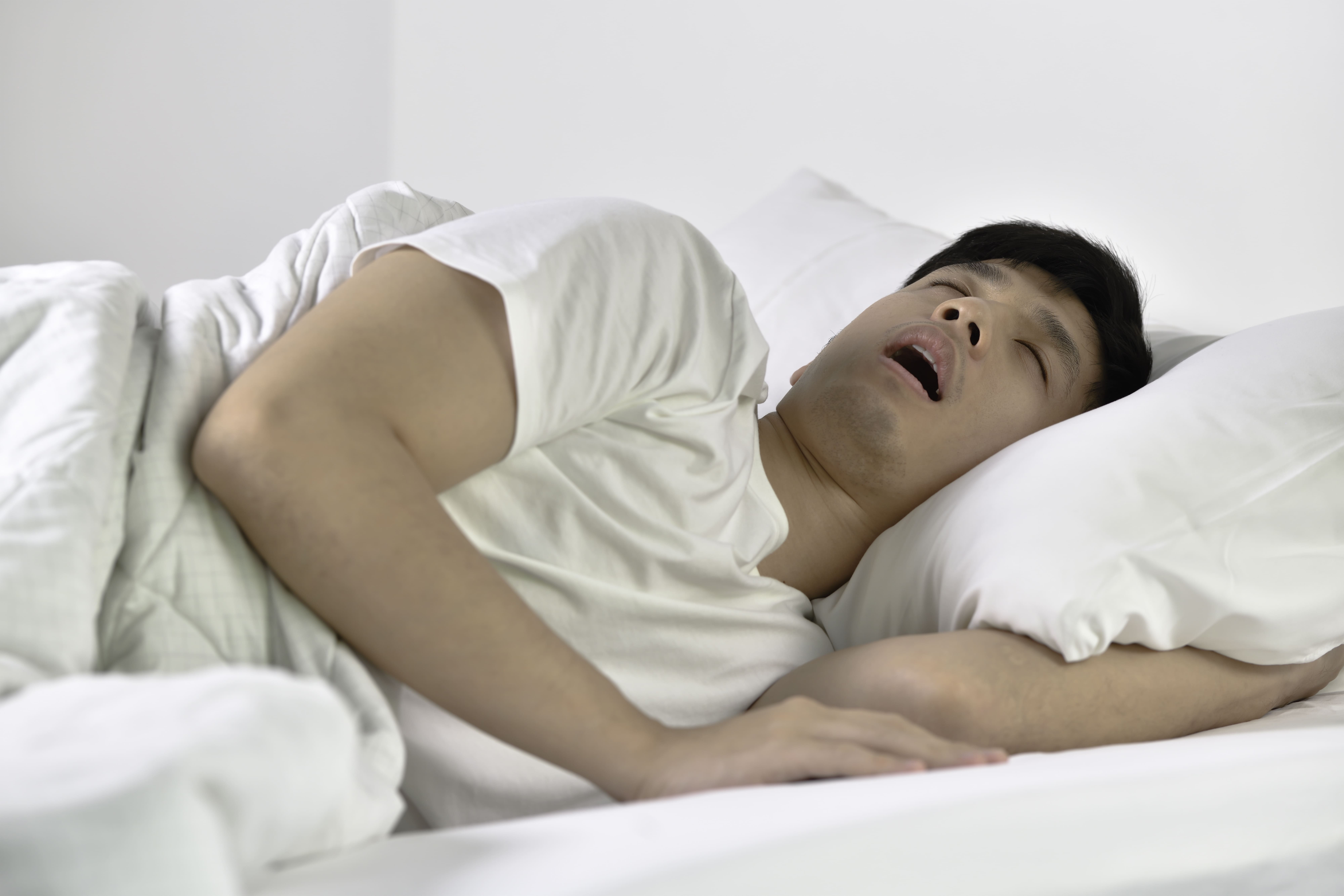
Does your partner report that you stop breathing during sleep? Sleep apnoea causes breathing interruptions during sleep, leading to fragmented sleep, reduced oxygen levels, and affecting heart health, cognitive function, and daily alertness. A sleep study is a diagnostic test used to identify sleep disorders by measuring breathing patterns, oxygen saturation, brain activity, and body movements throughout the night. This determines whether these interruptions occur and their severity, with data guiding treatment decisions from lifestyle modifications to CPAP therapy or surgical interventions.
Types of Sleep Studies Available
Polysomnography (PSG) – Laboratory Sleep Study
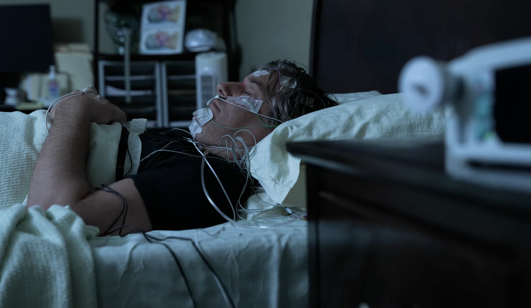
Polysomnography is a diagnostic tool for sleep apnoea conducted in a sleep laboratory with continuous monitoring by healthcare professionals. During PSG, sensors track multiple body functions:
- Brain waves are measured through EEG electrodes, which measure electrical activity in the brain
- Eye movements are detected via EOG sensors, which track how your eyes move during different sleep stages
- Muscle activity is recorded using EMG, which captures muscle tension and movement
- Heart rhythm is monitored with an ECG, which tracks your heart’s electrical signals
- Breathing effort is measured through chest and abdominal belts, which detect when your body tries to breathe
- Airflow is assessed via nasal cannulas, small tubes that measure air moving through your nose
- Oxygen levels are tracked through pulse oximetry, a finger clip that measures oxygen in your blood
- Leg movements are captured with additional sensors
The controlled laboratory environment allows technicians to adjust equipment throughout the night and document body positions. PSG captures all sleep stages. It distinguishes between obstructive events, in which the throat tissues block airflow despite breathing efforts, central events, in which the brain temporarily stops sending breathing signals, and mixed events that combine both patterns. This detailed analysis can identify not just sleep apnoea but also periodic limb movements (repeated leg twitches during sleep), REM sleep behaviour disorder (physically acting out dreams), and unusual arousal patterns that home studies might miss.
Laboratory studies may be considered for several patient groups:
- Patients with suspected central sleep apnoea
- Those with significant cardiac or pulmonary conditions
- Individuals who failed CPAP therapy
- Cases where initial home study results conflict with clinical symptoms
The data help specialists determine whether upper airway surgery might be helpful or whether combination therapies are needed.
Home Sleep Apnoea Test (HSAT)
Home sleep tests use portable monitoring devices that patients wear in their own beds. These devices measure:
- Breathing patterns
- Oxygen levels
- Heart rate
- Sometimes body position
A finger probe monitors oxygen levels, similar to the clip used in a doctor’s office. Chest and abdominal belts detect breathing effort by sensing when your chest and stomach move as you try to breathe. A nasal cannula measures airflow, a small tube that detects air moving through your nose. Some home units now include snoring microphones and position sensors that track whether apnoea events worsen when sleeping on the back.
Setup involves following the instructions to attach sensors correctly. Patients activate the recording before sleep and return the device for data download and analysis. Whilst home tests don’t measure sleep stages directly, they calculate the respiratory disturbance index (RDI), which counts breathing problems per hour by dividing the number of breathing events by the recording time.
Home testing may suit patients with a high pre-test probability of obstructive sleep apnoea based on symptoms such as loud snoring, witnessed breathing pauses, and daytime sleepiness, combined with risk factors such as obesity, a large neck circumference, or structural features such as enlarged tonsils. The convenience and lower cost make HSAT accessible for straightforward cases. However, negative results in symptomatic patients may require follow-up laboratory testing.
Watch-PAT and Wearable Devices
Watch-PAT technology uses peripheral arterial tone signals (changes in blood vessel activity in your fingertips), combined with pulse oximetry (oxygen level monitoring) and actigraphy (movement tracking), to detect sleep apnoea events through changes in vascular activity. The device is worn like a wristwatch with a finger probe. It identifies respiratory events by analysing arterial pulsation patterns, the rhythm of blood flow through small arteries, which change when breathing disruptions occur. This method differentiates sleep from wake periods without requiring EEG electrodes, making it less intrusive than traditional studies.
Consumer wearable devices and smartphone apps that claim to detect sleep apnoea use various combinations of movement tracking, sound recording, and optical sensors. Whilst these tools may identify snoring patterns or restless sleep, they cannot reliably diagnose sleep apnoea without measuring actual breathing patterns and oxygen levels. Their role remains limited to preliminary screening or tracking treatment response rather than definitive diagnosis.
What Happens During Assessment
Pre-Study Preparation
The preparation process begins with reviewing medications. Certain drugs affect sleep architecture or breathing patterns. Sedatives, muscle relaxants, and some cardiac medications may need adjustment. However, patients should not stop medications without medical consultation. Alcohol and caffeine restriction typically starts in the afternoon of the study day. Both substances alter sleep quality and breathing patterns.
For laboratory studies, patients bring comfortable sleepwear, their usual pillows if preferred, and any nightly medications. Technicians need access to the scalp to place small sensors that monitor brain activity. Recently washed, product-free hair facilitates sensor adhesion. Patients with CPAP machines bring them along if the study aims to assess treatment effectiveness or titrate pressure settings.
Home study preparation involves checking device batteries, understanding indicator lights, and practising sensor placement before bedtime. Instruction materials with diagrams show positioning for chest belts, finger probes, and nasal sensors. Many units include backup supplies like extra batteries and adhesive strips in case sensors detach during sleep.
During the Sleep Study
Laboratory polysomnography begins with sensor application. Technicians attach each electrode using conductive paste and secure it with gauze and tape. Bio-calibration follows. Patients perform specific movements like blinking, looking left and right, flexing their feet, and breathing deeply to verify that all channels record properly. Patients then settle into bed. Technicians monitor from an adjacent room via video and intercom systems.
Throughout the night, technicians document sleep positions, adjust sensors if signals deteriorate, and respond to patient needs. They observe for leg movements, unusual behaviours, and breathing patterns visible on video. If severe sleep apnoea appears early in the study, some laboratories perform split-night protocols. This involves introducing CPAP therapy halfway through to begin pressure titration immediately.
Home studies rely on patients maintaining sensors throughout the night without technical support. Many devices tolerate some movement and position changes. They continue to record even if certain channels temporarily lose signal. Patients document their bedtime, approximate sleep time, and any issues encountered. Morning removal follows specific sequences to preserve data before returning equipment.
Data Collection Parameters
Sleep studies generate physiological data. Polysomnography records multiple channels simultaneously. The apnoea-hypopnoea index (AHI) represents breathing events per hour of sleep. The oxygen desaturation index tracks drops from baseline oxygen levels in the blood. Minimum oxygen saturation identifies the lowest levels reached during breathing events.
Respiratory effort-related arousals (RERAs) capture breathing difficulties causing brief awakenings without meeting full apnoea or hypopnoea criteria. Technicians detect these events through oesophageal pressure monitoring or analysis of airflow patterns. These events can explain symptoms in patients with upper airway resistance syndrome who show normal AHI values but still experience sleep fragmentation.
Body position data reveals whether apnoea severity changes between back, side, or stomach sleeping. REM sleep analysis identifies whether breathing problems concentrate during dream sleep when muscle tone decreases. Heart rate variability patterns can suggest cardiovascular stress levels. Leg movement indices identify concurrent restless legs syndrome or periodic limb movement disorder requiring separate treatment.
Interpreting Results
Understanding Your Sleep Study Report
Sleep study reports begin with summary statistics. These include total sleep time, sleep efficiency (percentage of time asleep whilst in bed), and sleep stage distribution showing light sleep, deep sleep, and REM proportions. Normal adults spend a small portion in stage N1 (light sleep), approximately half in N2, a moderate amount in N3 (deep sleep), and roughly a quarter in REM sleep. These proportions vary with age and individual differences.
The respiratory event summary details different breathing disruption types:
- Obstructive apnoeas show ceased airflow despite continued chest and abdominal movement (meaning the person tries to breathe, but air cannot pass through)
- Central apnoeas lack both airflow and breathing effort (the brain temporarily stops sending signals to breathe)
- Hypopnoeas represent partial airflow reductions of a significant degree accompanied by oxygen desaturation (drops in blood oxygen levels) or arousal (brief awakening)
- Mixed events begin as central apnoeas then transition to obstructive patterns
The report specifies event duration, typically ranging from brief periods to over a minute, and their distribution across sleep stages and positions.
Oxygen saturation graphs display continuous overnight levels. They highlight desaturation episodes (drops in oxygen) corresponding to breathing events. Time spent below normal saturation indicates tissue hypoxia (insufficient oxygen reaching body tissues) severity. Prolonged periods suggest greater cardiovascular strain. The arousal index counts brief awakenings per hour, including both respiratory and non-respiratory causes. Frequent arousals can explain daytime fatigue even in mild sleep apnoea cases.
Severity Classifications
AHI values provide the primary severity classification, but a comprehensive assessment considers multiple factors. Your healthcare provider may interpret these results based on your specific symptoms, health history, and individual risk factors. Patients with mild-range AHI but significant oxygen desaturations face different risks than those with higher AHI but maintained oxygen levels. Similarly, someone with positional apnoea (breathing events that occur mainly in certain sleep positions) showing higher AHI when supine but lower AHI when sleeping laterally has different treatment options than someone with consistent apnoea regardless of position.
The oxygen desaturation index (which measures how often oxygen levels drop during sleep) often correlates with AHI but may diverge in certain patterns. Some patients show frequent brief apnoeas causing minimal desaturation. Others experience longer events with substantial oxygen drops. The cumulative oxygen burden calculates the total time and depth below normal saturation. It provides valuable information about cardiovascular consequences beyond event counts alone.
Symptom correlation can strengthen diagnostic certainty. Patients with moderate AHI but significant daytime dysfunction (such as fatigue, difficulty concentrating, or falling asleep during daily activities) may need treatment. Asymptomatic individuals discovered during screening might start with conservative measures. Your doctor may set treatment goals based on both your test results and how symptoms affect your daily life. The Epworth Sleepiness Scale score, documenting drowsiness likelihood in various situations, helps quantify functional impact beyond pure numbers.
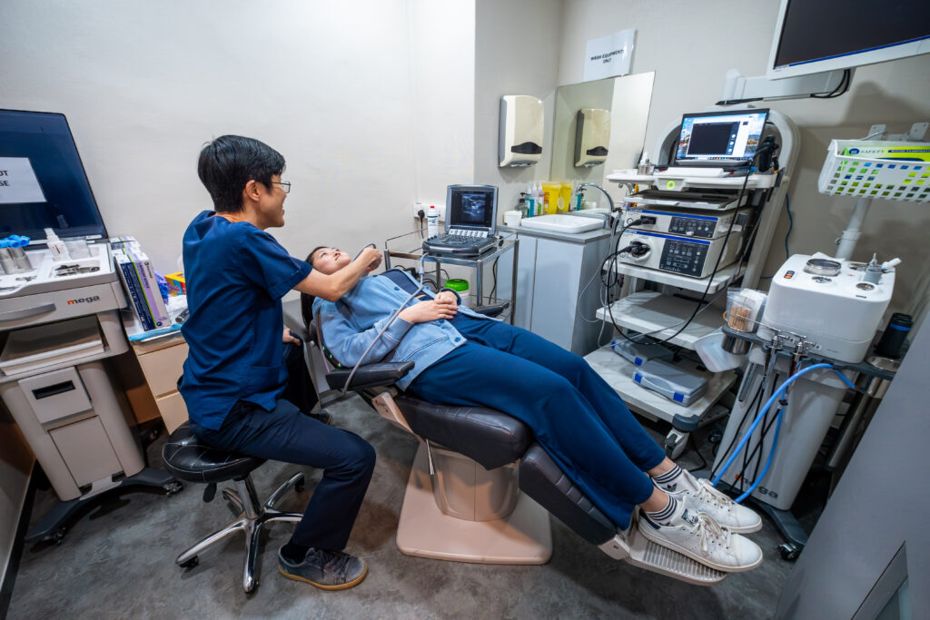
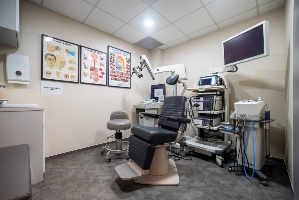
ENT Evaluation Components
Physical Examination
ENT specialists perform systematic upper airway examination, identifying anatomical factors contributing to airway collapse. Nasal assessment includes examining septum position (the wall dividing the nostrils), turbinate size (the structures inside the nose that warm and humidify air), and valve competence (how well the nasal opening maintains airflow). Nasal obstruction increases negative pressure during inspiration, promoting pharyngeal collapse. For many patients, successful sleep apnoea management requires addressing nasal resistance. Structural issues, such as a crooked midline, can make breathing difficult during the night; hence, Deviated Septum Surgery in Singapore may be recommended to improve nasal airflow and enhance the effectiveness of other sleep treatments. Flexible nasopharyngoscopy (a thin camera passed through the nose) visualises the entire airway from nostrils to larynx (voice box), revealing polyps (growths), adenoid tissue, or anatomical variants.
Determining the severity of Obstructive Sleep Apnoea (OSA) requires a combination of clinical history and objective data. Consulting a Sleep Specialist in Singapore is the first step toward performing a Polysomnogram (PSG) to monitor breathing patterns, oxygen levels, and heart rate during sleep.
Oral cavity examination evaluates tonsil size using standardised grading (absent to touching midline), tongue volume relative to oral space, and palate length and thickness. The modified Mallampati score assesses pharyngeal crowding by the visibility of structures with the mouth open and the tongue protruded. Retrognathia (recessed jaw) or micrognathia (small jaw) affect tongue position and posterior airway space. Specialists identify these through profile examination and dental occlusion assessment (how the teeth meet when the jaw closes).
The Müller manoeuvre, performed during flexible endoscopy, involves attempted inspiration against a closed nose and mouth. This creates negative pressure that simulates airway collapse during sleep. This dynamic assessment shows collapse levels at the soft palate, lateral pharyngeal walls, tongue base, or epiglottis (the flap covering the windpipe). However, awake findings don’t always predict sleep patterns. Some specialists use drug-induced sleep endoscopy for more accurate collapse pattern identification in surgical candidates.
Additional Diagnostic Tests
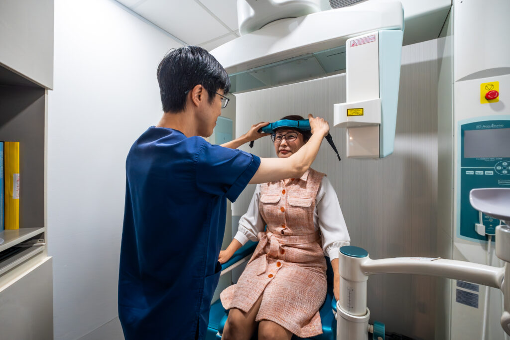
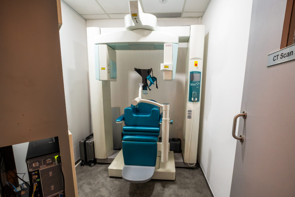
Lateral cephalometric X-rays measure skeletal relationships and soft tissue dimensions, quantifying posterior airway space, tongue size, and hyoid bone position (the U-shaped bone in the neck that supports the tongue). These measurements identify patients who might benefit from maxillomandibular advancement surgery (a procedure that repositions the jaw bones forward) rather than soft tissue procedures alone. Cone beam CT provides three-dimensional airway imaging, calculating minimum cross-sectional areas and identifying the narrowest segments.
Acoustic rhinometry and rhinomanometry measure nasal airway dimensions and resistance. These tests document obstruction severity and response to decongestants or positioning changes. They guide decisions about septoplasty (surgery to straighten the nasal septum), turbinate reduction (surgery to shrink nasal structures), or nasal valve surgery as adjuncts to sleep apnoea treatment. Peak nasal inspiratory flow offers a screening method that patients can perform at home.
For selected cases, sleep MRI captures dynamic airway changes during natural sleep without radiation exposure. It shows real-time collapse patterns and tissue vibration. This imaging helps predict surgical outcomes and identify multi-level obstruction requiring combination procedures. However, availability remains limited and costs substantial compared to standard assessments.
Treatment Planning Steps
Based on a comprehensive assessment combining sleep study data, physical examination findings, and patient factors, treatment proceeds through stepped approaches tailored to individual needs.
Mild sleep apnoea with positional components might start with:
- Weight management
- Side-sleeping devices
- Treatment of nasal obstruction (such as a blocked or stuffy nose) to improve airflow
Moderate to severe cases typically require CPAP therapy as first-line treatment. Mask fitting and pressure titration (adjustment to find the right air pressure level) for comfort and effectiveness.
When CPAP proves intolerable despite proper fitting and pressure adjustment, oral appliances (devices worn in the mouth) that advance the mandible (move the lower jaw forward) offer an alternative for mild to moderate cases. Dentists with sleep medicine training fabricate custom devices. These require titration (gradual adjustment) to balance effectiveness against jaw discomfort. In paediatric cases, sleep apnoea is frequently caused by physical obstructions in the throat or nasopharynx. If a sleep study confirms significant obstruction in a child, a consultant may discuss Adenoidectomy Surgery in Singapore as a primary treatment to clear the airway and restore healthy sleep cycles. Regular follow-up with repeat sleep studies confirms treatment response.
Surgical options address specific anatomical abnormalities (structural differences in your airway) when conservative measures fail, or patient preference favours definitive correction. Procedures range from minimally invasive radiofrequency ablation (using heat energy to shrink tissue) of the soft palate to skeletal surgery advancing both jaws (moving the upper and lower jaw bones forward). Multi-level surgery combining nasal, palatal (roof of mouth), and tongue base procedures may be necessary for complex obstruction patterns. Outcomes vary among patients based on collapse patterns, BMI (body mass index, a measure of body fat based on height and weight), and severity factors identified during evaluation.
Preparation Steps for Your Sleep Study
- Schedule strategically: Book your study during a typical work week rather than weekends when sleep patterns might differ. Avoid times of unusual stress or schedule disruptions that affect sleep quality.
- Maintain a regular sleep schedule: Keep consistent bedtimes and wake times for at least a week before the study. Irregular patterns can affect sleep architecture and study accuracy.
- Document symptoms: Record specific observations about breathing and sleep issues. Note snoring patterns, breathing pauses witnessed by partners, morning headaches, and daytime fatigue levels.
- Prepare practical items: For lab studies, pack loose-fitting sleepwear with button fronts allowing wire access. Bring personal hygiene items and any regular medications in original containers.
- Plan transportation: Arrange rides for laboratory studies. Morning grogginess after disrupted sleep and residual electrode paste in hair may make driving less comfortable.
When to Seek Professional Help
- Loud, persistent snoring that disturbs partners or occurs most nights
- Witnessed breathing pauses or gasping during sleep
- Morning headaches occurring several times weekly
- Falling asleep inappropriately during meetings, driving, or conversations (a sign of excessive daytime sleepiness)
- Unrefreshing sleep despite adequate time in bed
- Difficulty concentrating or memory problems developing gradually
- Blood pressure remains elevated despite medication
- Irregular heartbeat or palpitations (a sensation of your heart racing or skipping beats) during rest
- Night sweats unrelated to room temperature
- Frequent nighttime urination beyond typical patterns
Commonly Asked Questions
How accurate are home sleep studies compared to lab studies?
Home sleep studies can diagnose moderate to severe obstructive sleep apnoea in most cases. They have diagnostic agreement compared to polysomnography (a comprehensive sleep test conducted in a lab that monitors brain waves, breathing, and body movements). However, they may underestimate the severity since they measure recording time rather than actual sleep time. They cannot detect sleep stages or identify central apnoeas (breathing pauses caused by the brain not sending breathing signals), periodic limb movements, or other sleep disorders.
Can I use sleeping pills the night of my sleep study?
Avoid sleeping medications unless a sleep physician approves them, as they alter sleep architecture (the natural pattern of sleep stages your body cycles through) and may worsen breathing problems. If you regularly use prescribed sleep aids, discuss timing with the sleep centre beforehand. Natural sleep provides an assessment of your typical breathing patterns and sleep quality.
Will insurance cover my sleep study?
Sleep studies may be covered when medically indicated (when your symptoms suggest a sleep disorder that needs diagnosis), though pre-authorisation requirements vary. Laboratory polysomnography typically requires documentation of symptoms and failed conservative measures. Home studies often have less stringent criteria. Check coverage details, including deductibles and whether the sleep centre participates in your network.
How soon after the study will I get results?
Sleep study data requires analysis by healthcare professionals. Complete interpretation typically takes several business days. A sleep physician, a doctor who specialises in sleep disorders, reviews raw data, generates the formal report, and develops treatment recommendations before your follow-up appointment. Severe findings prompting urgent treatment may generate faster communication.
What if I can’t fall asleep during the study?
Sleep laboratories expect some difficulty given the unfamiliar environment and attached sensors. Even a few hours of sleep can often provide sufficient data for diagnosis, particularly if breathing abnormalities (such as pauses in breathing, gasping, or snoring) are consistently present. If absolutely no sleep occurs, the study may need to be repeated. This happens rarely with professionals creating comfortable conditions.
Conclusion
Sleep study results confirm sleep apnoea, guide treatment selection, and identify anatomical factors requiring intervention. Treatment effectiveness depends on proper CPAP therapy, positional modifications, or surgical correction based on individual obstruction patterns. Regular follow-up helps with optimal outcomes through monitoring and adjustments.
If you are experiencing loud snoring, breathing pauses, or excessive daytime fatigue, schedule a consultation with an ENT specialist, who can assess your symptoms, evaluate your upper airway anatomy, and recommend the appropriate sleep study assessment and management plan.