Nasal Endoscopy: What to Expect During the Assessment
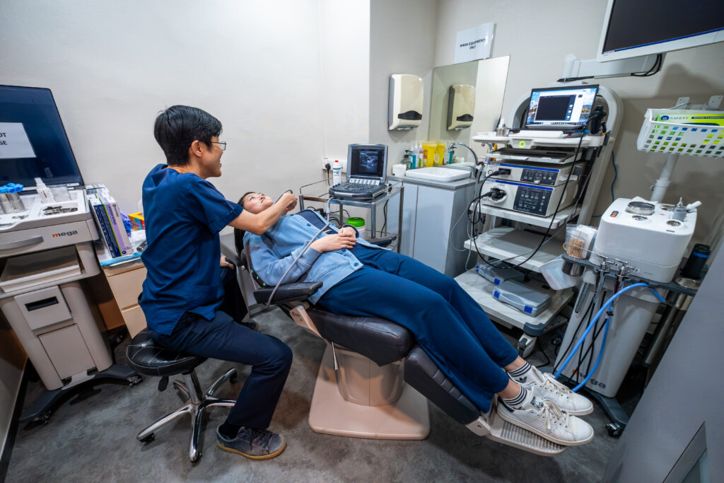
A nasal endoscopy can reveal polyps hidden deep in sinus cavities, structural abnormalities causing breathing difficulties, and early signs of inflammation, all invisible to standard examination. Your ENT specialist performs this assessment using a thin, flexible tube with a camera attached to examine the internal structures of your nose and sinuses. The procedure typically takes 1 to 5 minutes and provides immediate visual information that guides diagnosis and treatment planning.
Unlike imaging studies that show static pictures, nasal endoscopy offers real-time visualisation of moving structures. Your specialist can observe how your nasal passages function during breathing, identify areas of mucus pooling, and assess the health of tissues lining your sinuses. This direct observation complements CT imaging by providing mucosal detail and real-time functional information that static scans cannot capture.
How Nasal Endoscopy Works
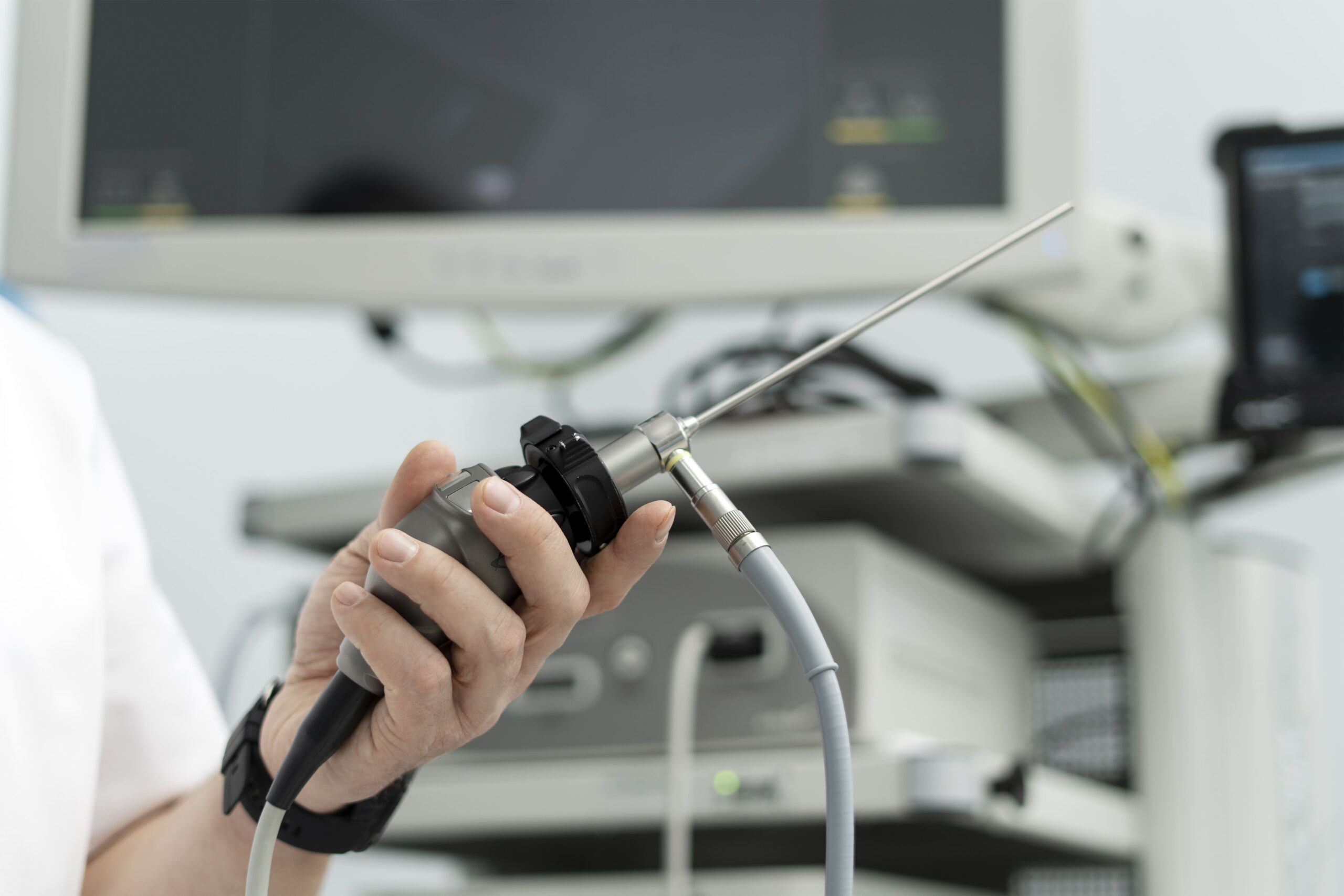
The endoscope used in nasal assessment measures approximately 2.7 to 4 millimetres in diameter—roughly the width of a drinking straw. It contains fibre-optic technology that transmits light and captures high-definition images. These images display on a monitor in real-time. It uses fibre-optic or digital imaging technology that transmits light and captures high-definition images displayed on a monitor in real-time. Rigid endoscopes, which are standard for diagnostic purposes, are advanced systematically through the nasal passages. Flexible endoscopes may also be used in certain clinical situations.
Modern endoscopes feature angled lenses, typically 0-degree, 30-degree, and 70-degree options. These allow visualisation around anatomical curves without repositioning the instrument. The 0-degree scope provides direct forward views, while angled scopes reveal spaces behind structures like the middle turbinate and within sinus openings.
Areas Examined During the Procedure
The examination follows a systematic pathway through your nasal anatomy. Starting at the nasal vestibule (the entrance), the scope advances along the floor of the nose toward the nasopharynx, the space behind your nose connecting to your throat. Along this path, your specialist examines:
- The inferior turbinate, a long shelf-like structure that warms, humidifies, and filters air, often swells in response to allergies or infection.
- The middle meatus, the drainage pathway for the frontal, maxillary, and anterior ethmoid sinuses, commonly harbours polyps or shows signs of chronic inflammation.
- The sphenoethmoidal recess, located superoposterior to the superior turbinate, is where the sphenoid sinus drains into the nasal cavity. The adjacent superior meatus receives drainage from the posterior ethmoid sinuses.
- The adenoid pad, visible in the nasopharynx, may enlarge and obstruct breathing or contribute to recurrent infections.
- Eustachian tube openings (the passages that connect your middle ear to the back of your throat and help regulate ear pressure), located on either side of the nasopharynx, can show signs of dysfunction affecting ear pressure regulation.
Preparing for Your Assessment
Follow your specialist’s specific instructions regarding nasal sprays before your appointment. Some clinicians prefer patients to avoid self-applying nasal decongestants prior to arrival, as a topical decongestant is typically administered by the specialist immediately before the procedure to widen the passages for easier navigation.
You may be asked to remove nasal piercings before the procedure. Inform your specialist about blood-thinning medications, recent nosebleeds, or any history of fainting during medical procedures.
What Happens Before the Scope Is Inserted
Your specialist first examines your nose externally, assessing symmetry and any visible deformity. A headlight examination using a speculum provides initial views of the anterior nasal cavity.
Before endoscopy, a topical anaesthetic spray numbs the nasal lining. This contains lidocaine (a medication that temporarily blocks pain signals in the tissue it touches), which typically takes effect within 2 to 5 minutes of application, though clinicians commonly allow up to 10 minutes before proceeding. Some practitioners add a decongestant to the spray, widening passages for easier navigation. The numbing sensation feels unusual, like when a dental anaesthetic affects your lip, but should not be painful.
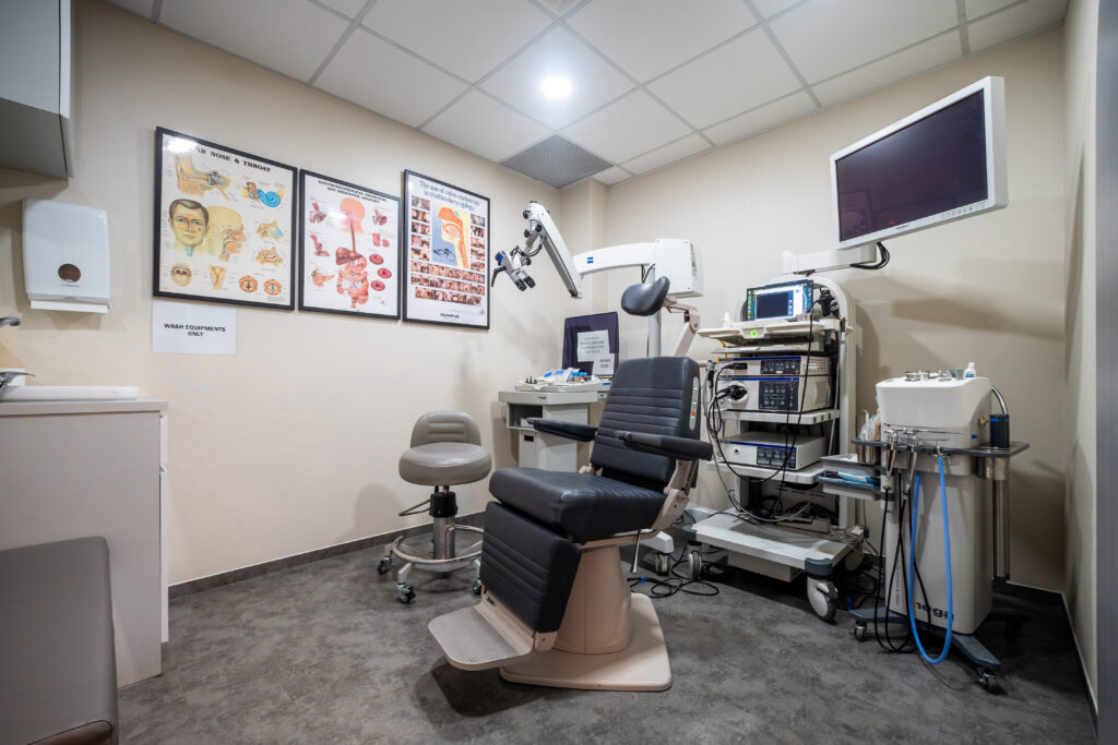
You’ll sit upright in an examination chair, head supported. Breathing through your mouth throughout the procedure keeps the scope pathway clear. Your specialist may ask you to swallow or say certain sounds to observe specific structures in motion.
During the Examination
The scope enters one nostril and advances slowly. You may feel pressure or slight discomfort, but it should not be painful. Brief discomfort occurs when passing narrow areas or touching sensitive spots. This triggers a momentary urge to sneeze or causes eyes to water reflexively.
Communication continues throughout. Your specialist describes findings as they appear on the monitor. They often turn the screen so you can observe. Seeing your own anatomy helps you understand explanations about your condition. Questions are welcome, though speaking may shift structures slightly.
💡 Did You Know?
The nasal cavity contains erectile tissue similar to that found elsewhere in the body. This tissue swells and shrinks in alternating cycles throughout the day, a phenomenon called the nasal cycle. One side typically dominates airflow while the other rests, switching every few hours.
Common Findings and What They Indicate
A deviated septum appears as a bend, shift, or spur in the central partition dividing the nose. Mild deviations rarely cause symptoms. Significant ones can obstruct airflow on one or both sides and may also contribute to recurrent sinus infections or nosebleeds.
Turbinate hypertrophy presents as enlarged, boggy (soft and swollen) tissue that narrows the breathing passages. This commonly responds to allergies, infection, or chronic irritation and often improves with medical treatment, such as nasal steroid sprays or saline rinses, before surgery becomes necessary.
Nasal polyps (soft, non-cancerous growths) appear as pale grey or yellowish, grape-like growths typically emerging from the middle meatus or sphenoethmoid recess. Unlike the surrounding pink, vascular nasal lining, polyps look semi-translucent and are mobile when assessed by the specialist. Their presence indicates chronic inflammation and typically requires ongoing medical management.
Mucopurulent discharge, thick, yellow-green mucus, may be associated with infection, though colour alone does not reliably distinguish a bacterial infection from a viral one. Your specialist will consider the discharge in context with your other symptoms and examination findings. Clear, excessive mucus more commonly suggests allergic or vasomotor rhinitis. The location where discharge is seen during endoscopy helps identify which sinuses may be involved.
After the Procedure
The numbing effect typically wears off within 20 to 30 minutes. If no biopsy was taken, you may blow your nose gently. Avoid eating or drinking until the numbness in your mouth and throat has fully resolved. Small amounts of blood-tinged mucus are common and not a cause for concern. You can resume normal activities immediately; no recovery period is necessary.
Some people experience mild headache or nasal irritation lasting a few hours. Over-the-counter pain relief may help manage this if needed. Contact your clinic if you develop significant bleeding, fever, or worsening symptoms over the following days.
Understanding Your Results
Your specialist discusses findings immediately after the examination. They often use captured images or video to illustrate. Written reports accompany referrals or become part of your medical record for future comparison.
Results fall into categories:
- Normal anatomy with no abnormalities
- Findings explaining your symptoms
- Incidental discoveries unrelated to your concerns
- Conditions requiring further investigation with imaging or biopsy (the doctor removes a small tissue sample for laboratory analysis)
Normal findings provide reassurance but don’t always explain symptoms completely. Functional issues, how structures behave during breathing or in response to triggers, sometimes require additional testing beyond visual examination. Your doctor will determine the appropriate next steps based on your specific situation and symptoms.
Conditions Diagnosed Through Nasal Endoscopy
Chronic rhinosinusitis (long-term inflammation of the nasal passages and sinuses) shows characteristic signs: polyps, thickened mucosa, and purulent discharge from sinus openings. The endoscopic appearance often correlates with symptom severity. It helps determine whether medical or surgical treatment suits your situation.
Cerebrospinal fluid leaks present as persistent, clear, watery discharge — often from one side of the nose. Nasal endoscopy may help identify the site of leakage, though confirmatory tests such as beta-2 transferrin analysis and CT imaging are typically required. Many CSF leaks resolve with conservative management; surgical repair via endoscopic techniques is performed when conservative measures fail.
Nasal tumours. It is worth noting that polyps or masses appearing on only one side of the nasal cavity warrant particular attention, as unilateral growths may require investigation to rule out malignancy.
Foreign bodies, particularly in children, lodge in the nasal passages and cause one-sided symptoms. Endoscopy locates these objects. It often enables removal during the same examination.
When Repeat Endoscopy Is Recommended
Monitoring chronic conditions may require periodic reassessment. Nasal polyps recur frequently despite treatment. Regular endoscopy detects regrowth before symptoms worsen. Post-surgical patients undergo scheduled examinations to ensure proper healing and identify complications early.
Changing symptoms warrant repeat examination. New obstruction, altered discharge characteristics, or development of facial pain may suggest disease progression. Note that severe or worsening facial pain, particularly alongside one-sided symptoms, should prompt ENT review rather than routine reassessment.
Practical Steps for Your Appointment
Before arriving: Note your symptoms’ duration, triggers, and what makes them better or worse. List all nasal medications, including over-the-counter sprays you’ve tried.
During the procedure: Breathe steadily through your mouth. Alert your specialist if you feel faint or need a break. Though the examination is brief, your comfort matters.
After leaving: Write down any instructions while fresh in your mind. Photograph prescription details if handwriting proves difficult to read later.
Avoid eating or drinking until any numbness in your mouth or throat has fully resolved, typically within 30 to 60 minutes. Write down any instructions while fresh in your mind and photograph prescription details if handwriting proves difficult to read later.
When to Seek Professional Help
Seek routine ENT evaluation for:
- Nasal obstruction persists beyond 8 to 12 weeks despite first-line treatment
- Recurrent sinus infections requiring multiple courses of antibiotics
- Loss of smell is not recovering after a recent cold
- Facial pressure or pain concentrated around the cheeks or forehead
- Post-nasal drip causing persistent throat clearing or cough, not responding to treatment
- Nosebleeds occurring frequently or without an obvious cause
Seek prompt ENT evaluation for:
- One-sided nasal symptoms, discharge, obstruction, or bleeding, particularly if persistent or worsening
- Severe facial pain that does not respond to over-the-counter pain relief
Commonly Asked Questions
Does nasal endoscopy hurt?
The anaesthetic spray may also temporarily numb your mouth and throat. Avoid eating or drinking until the numbness completely wears off, usually within 30 to 60 minutes after the procedure.
How long does the examination take?
The endoscopy itself typically takes between 1 and 5 minutes in total. Your full appointment, which includes history-taking, external examination, anaesthetic application, and discussion of findings, will generally take longer, though the scope examination itself is brief.
Can I drive home afterwards?
Yes. No sedation is used for diagnostic nasal endoscopy, so your vision, coordination, and alertness are unaffected. The topical anaesthetic may temporarily numb your nose, mouth, and throat, but this wears off within 30 to 60 minutes and does not impair driving.
Will I need a CT scan as well?
This depends on your symptoms and endoscopic findings. Endoscopy and CT imaging provide complementary information. Endoscopy shows mucosal detail and real-time function, while CT reveals bony anatomy and sinus contents. Your specialist can recommend imaging when findings suggest disease beyond what endoscopy visualises.
How often should nasal endoscopy be repeated?
Chronic rhinosinusitis with polyps, particularly for patients on biologic therapy or post-surgical follow-up, may require review every 3 to 6 months during active treatment. The appropriate interval depends on treatment type, disease severity, and your specialist’s clinical judgement.
Next Steps
Nasal endoscopy provides real-time visualisation that standard examination cannot. It identifies polyps, structural abnormalities, discharge patterns, and signs of chronic rhinosinusitis in a single procedure. When symptoms persist or recur, endoscopic findings directly determine whether treatment should be medical, surgical, or observational, making it a practical first step before committing to imaging or intervention.
If you are experiencing persistent nasal obstruction, recurrent sinus infections, loss of smell, one-sided nasal symptoms, or unexplained nosebleeds, consult an ENT specialist to discuss nasal endoscopy as part of your evaluation.