Maxillary Sinusitis: Everything You Need To Know

Did you know that the maxillary sinuses are the only paranasal sinuses present at birth, yet they’re the most prone to infection due to their unique drainage anatomy? Maxillary sinusitis occurs when the largest pair of sinuses, located in your cheekbones, become inflamed and filled with fluid. This condition causes facial pain, cheek pressure, and difficulty breathing through the nose. The maxillary sinuses connect to your nasal cavity through small openings called ostia, which can become blocked during inflammation, trapping mucus and bacteria inside.
The maxillary sinuses serve multiple functions: they lighten the skull weight, provide voice resonance, and produce mucus that moisturises nasal passages. When infection or inflammation affects these air-filled spaces, normal drainage stops, creating an environment in which bacteria or viruses thrive.
Anatomy and Function of Maxillary Sinuses
The maxillary sinuses sit within the cheekbones on either side of your nose, shaped like pyramids with their apex pointing toward the cheekbone. Each sinus measures approximately 15ml in volume and is lined with mucus-producing cells and tiny hair-like structures called cilia. These cilia beat in coordinated waves, moving mucus toward the ostium for drainage into the middle meatus of the nasal cavity.
The ostium’s location poses a challenge: it is high on the medial wall of the sinus, making gravity-assisted drainage difficult when standing or sitting. This anatomical feature explains why maxillary sinusitis often persists longer than infections in other sinuses. The sinus floor lies close to the roots of the upper teeth, particularly the molars and premolars, creating a pathway for dental infections to spread into the sinus.
Blood supply comes from branches of the maxillary artery, while sensory innervation arrives via the maxillary division of the trigeminal nerve. This nerve distribution explains why maxillary sinusitis often causes pain in the upper teeth, even when the teeth are healthy.
Causes and Risk Factors
Viral upper respiratory infections trigger most cases of acute maxillary sinusitis. Common cold viruses cause swelling of the nasal mucosa that extends into the sinus openings, thereby blocking normal drainage. Bacterial infection may follow, with Streptococcus pneumoniae, Haemophilus influenzae, and Moraxella catarrhalis being frequent culprits.
Dental infections, particularly of the upper molars and premolars, account for odontogenic maxillary sinusitis. Root canal infections, periodontal disease, or complications from dental procedures can introduce bacteria directly into the sinus through the thin bone separating tooth roots from the sinus floor.
Anatomical variations increase susceptibility to maxillary sinusitis:
- Deviated nasal septum blocking the ostium
Maxillary sinusitis can often be exacerbated by structural abnormalities within the nasal cavity. If a crooked midline is preventing proper drainage, a consultant may recommend Deviated Septum Surgery in Singapore to improve the nasal airway and facilitate better sinus health.
- Concha bullosa (air-filled middle turbinate) compresses drainage pathways
- Haller cells (ethmoid air cells extending into the maxillary sinus roof)
- A narrow infundibulum reduces drainage capacity
Environmental factors contribute through different mechanisms. Allergens cause mucosal swelling and excessive mucus production. Air pollution and cigarette smoke paralyse cilia function, impairing mucus clearance. Swimming pool chlorine irritates sinus linings, while air travel pressure changes can force nasal secretions into sinuses.
💡 Did You Know?
The maxillary sinus is the only paranasal sinus present at birth, though it remains small until permanent teeth develop. The sinus expands downward as adult teeth emerge, reaching full size around age 18.
Symptoms and Clinical Presentation
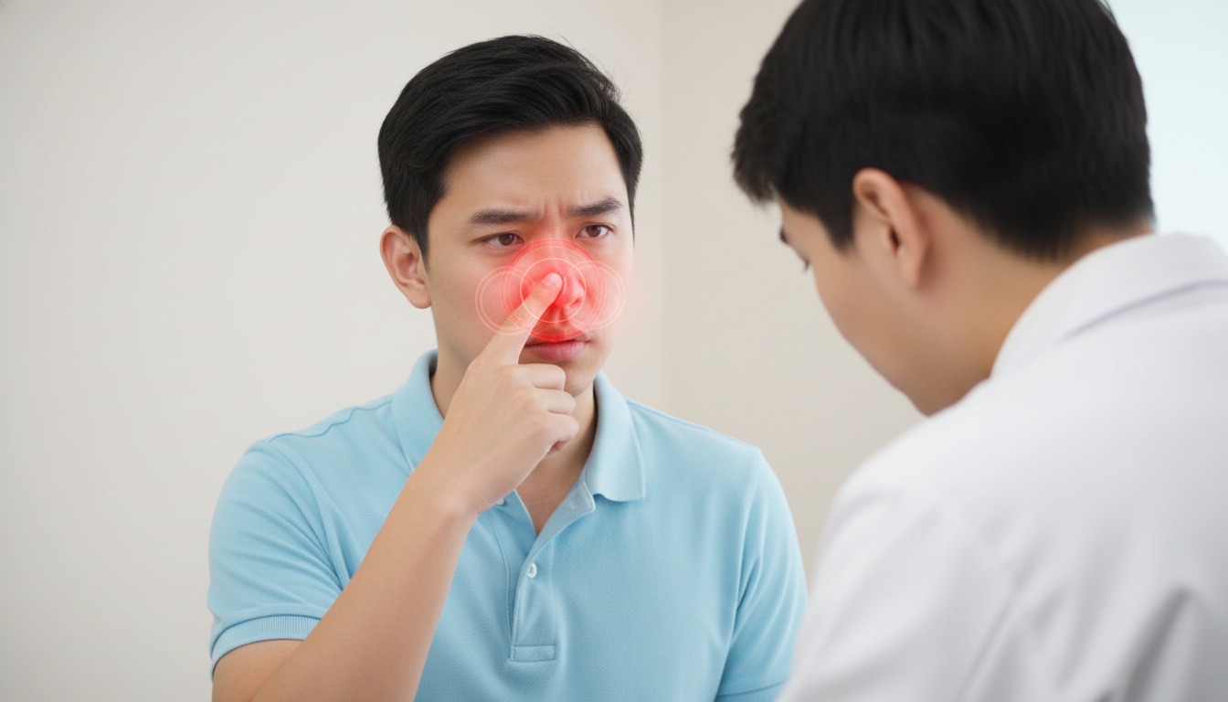
Facial pain and pressure characterise maxillary sinusitis, typically worsening when bending forward or lying down. Pain concentrates over the cheekbones and may radiate to the upper teeth, forehead, or temple. Patients may report that their upper teeth feel loose or aching, even in the absence of dental pathology.
Nasal symptoms include:
- Thick, discoloured nasal discharge (yellow, green, or blood-tinged)
- Nasal congestion, often unilateral
- Reduced or altered sense of smell
- Post-nasal drip is causing throat irritation
Systemic symptoms accompany bacterial infections: fever above 38°C, fatigue, and general malaise. Halitosis often develops from bacterial growth in stagnant mucus. Some patients experience ear fullness due to eustachian tube dysfunction caused by postnasal drip.
Chronic maxillary sinusitis presents with symptoms that persist for more than 12 weeks. Pain may be minimal or absent, replaced by persistent nasal congestion, post-nasal drip, and intermittent facial pressure. Nasal polyps sometimes develop, further obstructing drainage.
⚠️ Important Note
Severe headache with high fever, vision changes, or facial swelling requires immediate medical attention as these may indicate serious complications like orbital or intracranial extension of infection.
Diagnosis Methods
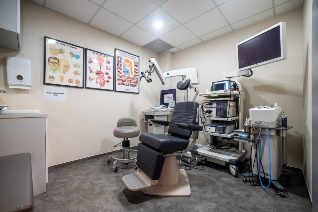
Physical examination begins with anterior rhinoscopy using a nasal speculum. ENT specialists look for mucosal swelling, purulent discharge from the middle meatus, and anatomical abnormalities. Transillumination – shining light through the sinuses – may show opacity in affected sinuses, though this technique has limited sensitivity.
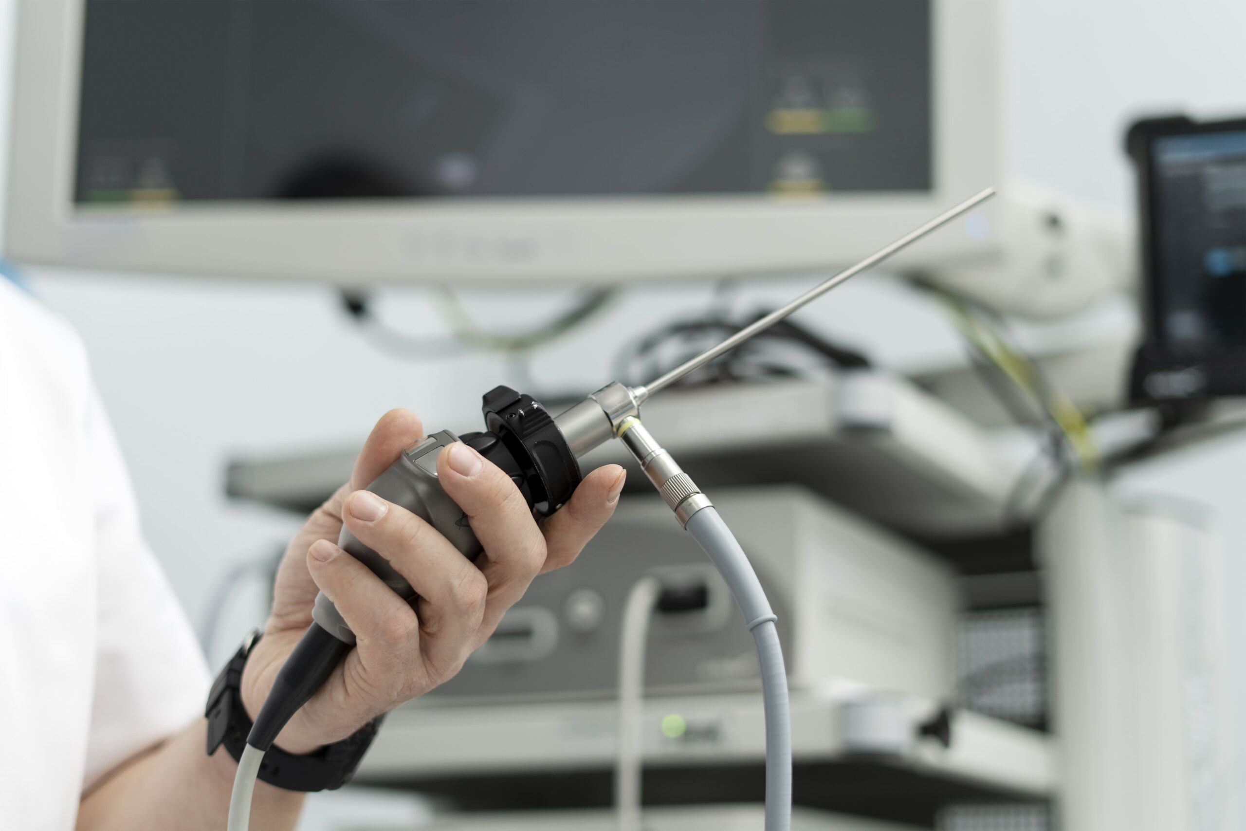
Nasal endoscopy provides detailed visualisation of the middle meatus and the drainage pathways of the sinuses. The procedure uses a thin, flexible tube with a camera to examine areas invisible to standard examination. Endoscopy reveals polyps, foreign bodies, tumours, and locations of purulent discharge.
Imaging studies confirm the diagnosis when clinical findings are unclear:
- X-rays show air-fluid levels or complete sinus opacification, but miss early changes
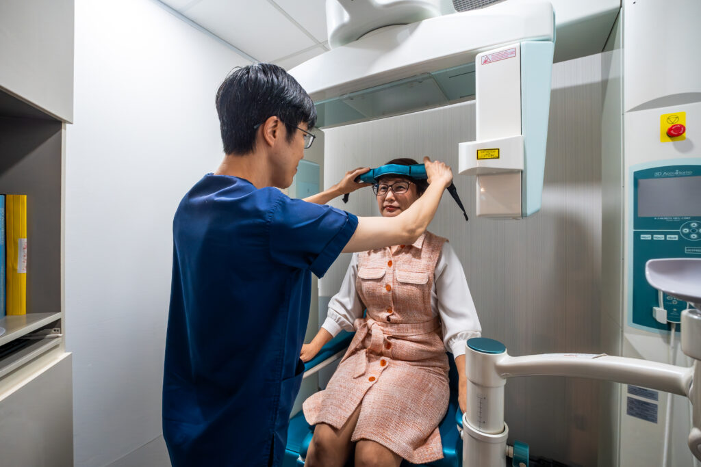
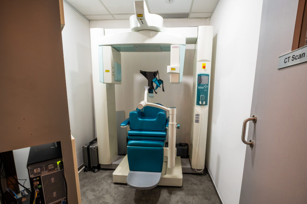
- CT scans provide detailed bone and soft tissue imaging, revealing the extent of disease
- MRI distinguishes between inflammation, fungal infections, and tumours
Maxillary sinus puncture and aspiration, though rarely performed now, provides a definitive bacteriological diagnosis. The procedure involves inserting a needle through the inferior meatus to obtain sinus contents for culture.
Treatment Approaches
Conservative Management
Saline nasal irrigation mechanically removes mucus, allergens, and inflammatory mediators. Isotonic saline (9g salt per litre) or hypertonic solutions may be used. The frequency and method of irrigation should be determined by a healthcare professional using squeeze bottles, neti pots, or powered devices.
Steam inhalation liquefies thick secretions, though evidence for significant benefit remains limited. Menthol or eucalyptus oil may be added for subjective symptom relief. Adequate hydration thins mucus naturally – appropriate fluid intake should be discussed with a healthcare professional, particularly if medical conditions are present.
Medical Treatment
Nasal corticosteroid sprays may be prescribed to help reduce inflammation in the nasal passages and shrink polyps. Correct spray technique is essential — the nozzle should be directed slightly outward toward the ear rather than upward. A healthcare professional should determine the type and duration of treatment.
Antibiotic therapy may be required if a bacterial infection is suspected. The choice of antibiotic, dosage, and treatment duration should be guided by a healthcare professional based on the individual’s condition and response to treatment.
Decongestant medications can provide temporary relief from nasal blockage. However, they should be used only as directed, as prolonged or unsupervised use — particularly of nasal sprays — may worsen congestion over time. Seek professional medical advice before starting or continuing decongestant therapy.
Surgical Intervention
When conservative treatments fail to resolve the inflammation, surgical intervention may be required to widen the sinus openings. Functional Endoscopic Sinus Surgery Singapore is a common procedure used to remove obstructive tissue and restore normal ventilation to the maxillary sinuses.
Functional endoscopic sinus surgery (FESS) may be considered when medical management is insufficient. The procedure enlarges the natural ostium, removes diseased tissue, and corrects anatomical abnormalities blocking drainage. Current techniques preserve healthy mucosa while establishing adequate ventilation.
Balloon sinuplasty offers a less invasive option for selected cases. A small balloon inflates within the blocked ostium, fracturing surrounding bone to widen the opening. The procedure preserves mucosa and may cause less post-operative discomfort.
For certain patients with maxillary sinus obstruction, a less invasive “keyhole” approach may be suitable. Consulting a Balloon Sinuplasty Specialist in Singapore allows you to explore options where a small balloon catheter is used to dilate the sinus ostia without the need for tissue removal.
Complications to Watch For
Orbital complications occur when infection spreads through the thin lamina papyracea separating the maxillary sinus from the orbit. Periorbital cellulitis typically presents with eyelid swelling and redness. Orbital cellulitis typically presents with proptosis, ophthalmoplegia, and vision changes that require urgent surgical drainage.
Intracranial spread carries serious consequences. Meningitis can develop via direct extension or hematogenous spread. Brain abscess and cavernous sinus thrombosis represent life-threatening complications requiring immediate neurosurgical consultation.
Chronic osteomyelitis of the maxilla develops from prolonged infection, particularly in immunocompromised patients. The condition causes bone destruction, facial deformity, and may require extensive surgical debridement.
✅ Quick Tip
Track your symptoms in a diary, noting triggers like weather changes, allergen exposure, or dental procedures. This information helps your healthcare professional identify patterns and determine appropriate treatment.
Putting This Into Practice
- Optimise your environment: Use HEPA filters in bedrooms and workspaces to reduce allergen exposure. Maintain indoor humidity at moderate levels by using humidifiers during dry seasons or by air conditioning during humid periods.
- Learn the nasal irrigation technique: Lean forward over a sink, turn your head to the side, and gently squeeze saline solution into the upper nostril. Allow drainage from the lower nostril without forceful blowing. Switch sides and repeat.
- Identify your triggers: Note activities, foods, or environments that precede episodes of sinusitis. Common triggers include dairy products, wine, perfumes, and weather fronts. Avoiding identified triggers may reduce the frequency of episodes.
- Maintain dental health: Schedule regular dental checkups, particularly for the upper teeth. Address cavities, gum disease, or dental infections promptly to prevent spread to the maxillary sinuses.
- Support your immune system: Regular exercise, adequate sleep, and stress management support immune function. Consult a healthcare professional regarding vitamin D supplementation if serum vitamin D levels are low, as deficiency is associated with increased risk of respiratory infections.
When to Seek Professional Help
- Facial pain or pressure lasting more than 10 days despite over-the-counter treatment
- High fever above 39°C with severe headache
- Vision changes, double vision, or eye swelling
- Facial swelling or redness extending beyond the cheek area
- Confusion or difficulty staying awake
- Neck stiffness with fever
- Multiple episodes of sinusitis within one year
- Blood-tinged nasal discharge persisting beyond several days
- Complete loss of smell lasting more than two weeks
- Dental pain affecting multiple upper teeth simultaneously
Commonly Asked Questions
Can maxillary sinusitis resolve without antibiotics?
Many cases of viral maxillary sinusitis resolve within 7-10 days with conservative measures such as saline irrigation, adequate rest, and hydration. Bacterial infections typically require antibiotics, especially when symptoms persist beyond 10 days or worsen after initial improvement.
Why does flying make my sinusitis worse?
Cabin pressure changes during ascent and descent affect sinus pressure equalisation. Blocked ostia prevent pressure equilibration, causing pain (barosinusitis). Using a decongestant nasal spray 30 minutes before descent may help maintain ostial patency. Consult a healthcare professional for appropriate guidance.
How do I differentiate between dental pain and sinus pain?
Sinus-related tooth pain typically affects multiple adjacent teeth, worsens with bending forward, and accompanies other sinus symptoms. Dental pain usually localises to one tooth, persists regardless of position, and may worsen with hot/cold stimuli or chewing.
Can chronic maxillary sinusitis cause permanent damage?
Untreated chronic sinusitis can lead to mucosal thickening, polyp formation, and bone remodelling. While these changes may be irreversible, appropriate treatment can prevent progression and manage symptoms.
Is maxillary sinusitis contagious?
The underlying viral infections causing acute sinusitis are contagious, but sinusitis itself isn’t directly transmissible. Good hand hygiene and avoiding close contact during acute illness reduce the risk of viral transmission.

Conclusion
Key management strategies include proper nasal irrigation technique, identification of personal triggers, and maintenance of good dental hygiene to prevent odontogenic infections. Early treatment with appropriate antibiotics or corticosteroids prevents complications like orbital cellulitis or chronic osteomyelitis. Surgical intervention may be necessary when anatomical abnormalities or failures of medical management occur.
If you are experiencing persistent facial pain, nasal congestion, thick nasal discharge, or recurrent sinus infections, an ENT specialist can provide a comprehensive evaluation and specialised treatment options.